How does the heart work?
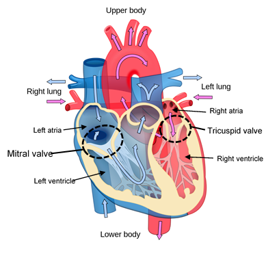
The heart consists of four chambers. The upper chambers are the right and left atria. The lower chambers are the right and left ventricles.
Blood flows from the body into the right atrium. It is then pumped into the right ventricle. The right ventricle pumps blood into the lungs, where it receives oxygen. It flows from the lungs into the left atrium; it is then is pumped into the left ventricle.
What is the mitral valve?
Each side of the heart has a valve that prevents blood from flowing backwards. The mitral valve is the valve separating the left atrium and the left ventricle. The tricuspid valve is the valve separating the right atrium and ventricle.
Because of the very large pressure created when the left ventricle contracts, the mitral valve can wear out in many dogs. This wearing out process begins with a small leak that gradually gets more severe. Disease of the tricuspid valve can occur as well, especially with progression of heart disease, though mitral valve disease is usually seen more commonly.
How does heart failure occur?
When the mitral valve leaks, it creates turbulence from the backflow of the blood. This can be heard as a heart murmur on auscultation and is usually the first sign noted of heart disease.
Over time, the leak can become more severe, causing more blood to backflow. This affects the pumping efficiency of the heart and leads to congestive heart failure.
When the mitral valve is affected, this increase of pressure on the left side of the heart causes fluid to leak into the tissues of the lungs. Increased of fluid in the lung tissues causes an accumulation of fluid in the lungs (pulmonary oedema) which leads to coughing, difficulty breathing and intolerance when exercising.
What tests are done to assess the situation?
There are several tests that are used to give the best evaluation of the heart function. Our veterinarian will explain and discuss these with you accordingly.
- Physical Examination and listening with a stethoscope (Auscultation) . Physical examination allows us to assess your pet for any abnormal findings, including their breathing pattern. Auscultation allows identification of murmurs, their location, and their intensity. It also allows us to hear lung sounds and if there is any fluid accumulation.
- Chest radiographs (X-rays). Chest radiographs are important to examine the size and shape of the heart. Radiographs will help us examine the lungs and if there is fluid accumulation present. Monitoring of these signs for any change is important in patients with heart disease.
- Ultrasound examination (Echocardiogram). Ultrasound examination allows us to examine the inside components of the heart in real time. This allows direct visualization of the valves and the other structures of the heart.
- Blood and urine tests. These tests allow detection of other disorders in the body that can impact on heart function such as kidney or liver disease. The knowledge of other organ function is also important in the treatment and response to medications.
- Electrocardiogram (ECG). This helps to assess the electrical activity of the heart to determine if any abnormal rhythms (arrhythmias) are present.
What treatment options are there?
A leaky heart valve can be replaced surgically in people. However, this is not as available in animals as of yet. Medical therapy and close monitoring is therefore crucial in these patients to manage their disease as best we can.
Several different drugs are used to treat and manage heart failure. These medications work in different ways with the ultimate aim to reduce the workload on the heart, so as to improve heart function. The results of the various tests will determine which medications are appropriate for your pet.
What can I do at home?
Close monitoring is an important step in determining the state and progression of heart disease. This, together with regular reviews, is important in ensuring the current medical regime is working for your pet or if it needs to be altered.
A good way to monitor at home is to monitor your pet’s “Sleeping/Resting Respiratory Rate (SRR/RRR)”. This is an excellent way to monitor heart disease as well as responses to medications. This should be started initially every 2-3 days in the initial phase followed by once-twice weekly.
Click here for our SRR instruction and monitoring sheet.